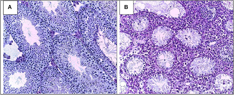
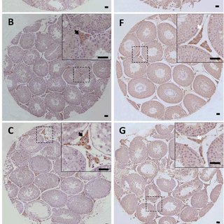
Morphology of Leydig cells in the testes after in vivo MCP-1 treatment.
Por um escritor misterioso
Last updated 03 junho 2024

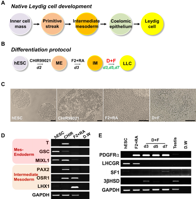
Rapid Differentiation of Human Embryonic Stem Cells into Testosterone-Producing Leydig Cell-Like Cells In vitro

Transcription factor Dmrt1 triggers the SPRY1-NF-κB pathway to maintain testicular immune homeostasis and male fertility
Frontiers Pathomechanisms of Autoimmune Based Testicular Inflammation
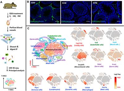
Frontiers Identification of Rat Testicular Leydig Precursor Cells by Single-Cell-RNA-Sequence Analysis
IJMS, Free Full-Text

Cell Type-Specific Expression of Testis Elevated Genes Based on Transcriptomics and Antibody-Based Proteomics

From Ancient to Emerging Infections: The Odyssey of Viruses in the Male Genital Tract
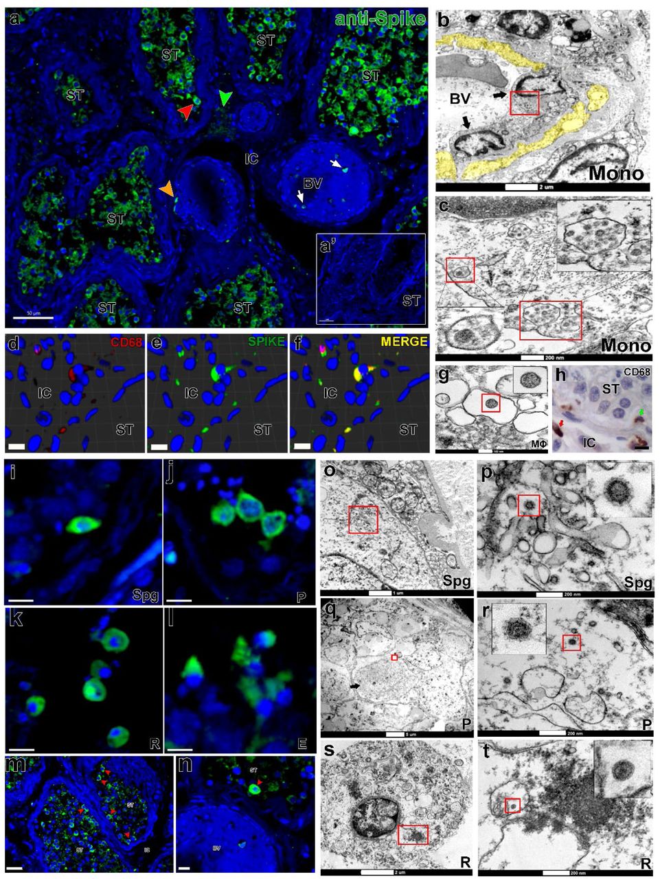
SARS-CoV-2 infects, replicates, elevates angiotensin II and activates immune cells in human testes

Can mesenchymal stem cells improve spermatogonial stem cell transplantation efficiency? - Kadam - 2017 - Andrology - Wiley Online Library
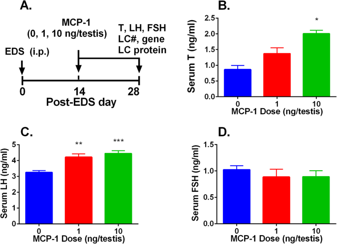
Monocyte Chemoattractant Protein-1 stimulates the differentiation of rat stem and progenitor Leydig cells during regeneration, BMC Developmental Biology
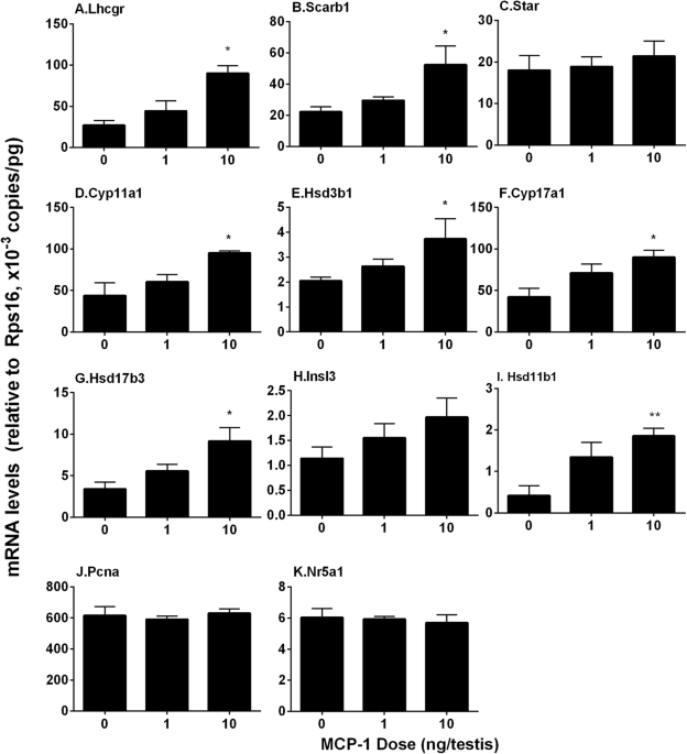
Monocyte Chemoattractant Protein-1 stimulates the differentiation of rat stem and progenitor Leydig cells during regeneration, BMC Developmental Biology

Transcription factor Dmrt1 triggers the SPRY1-NF-κB pathway to maintain testicular immune homeostasis and male fertility

Morphology of Leydig cells in the testes after in vivo PTHrP
PDF) Monocyte Chemoattractant Protein-1 stimulates the differentiation of rat stem and progenitor Leydig cells during regeneration
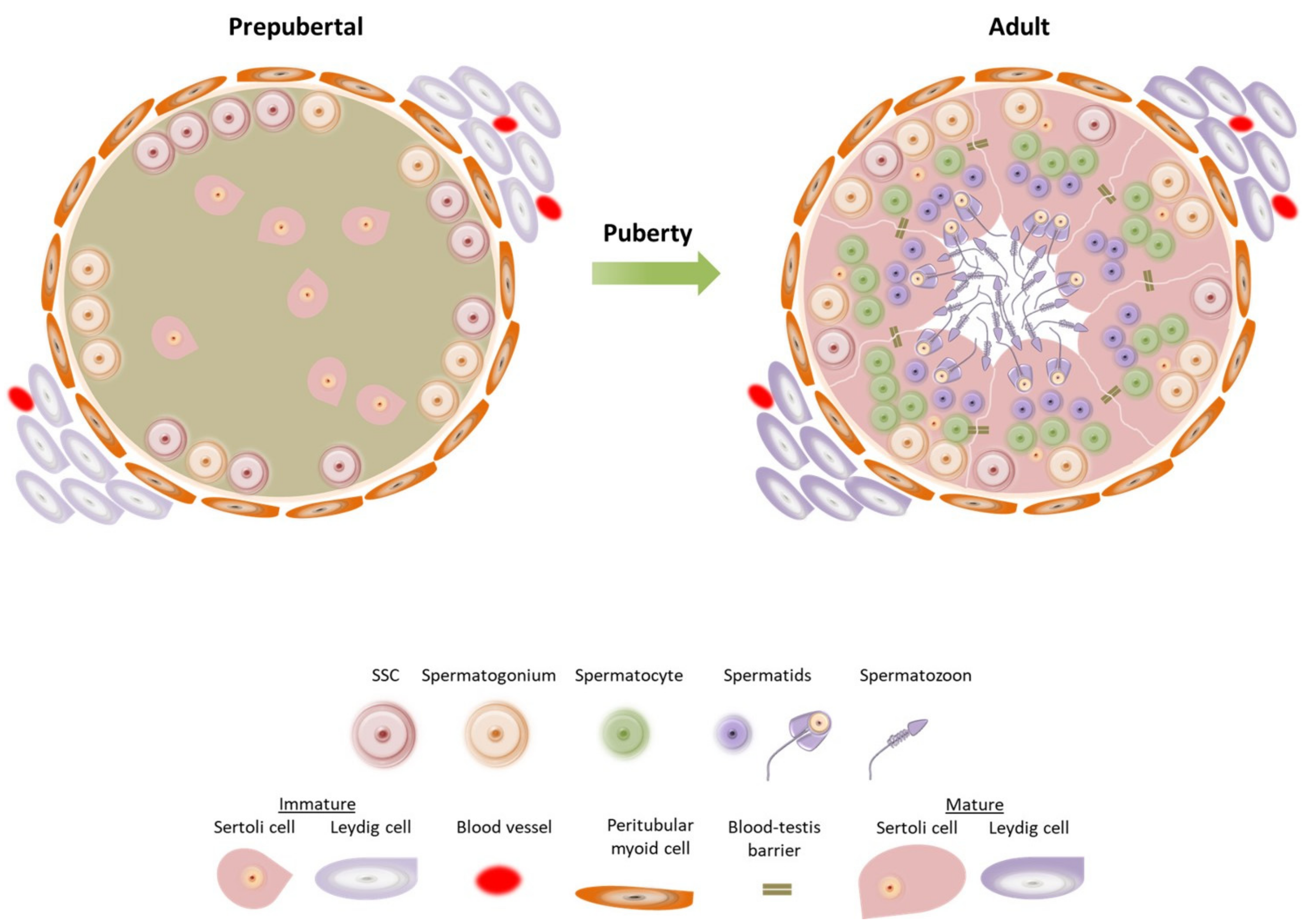
Sertoli cell ablation in adulthood induces apoptotic loss of Leydig
Recomendado para você
-
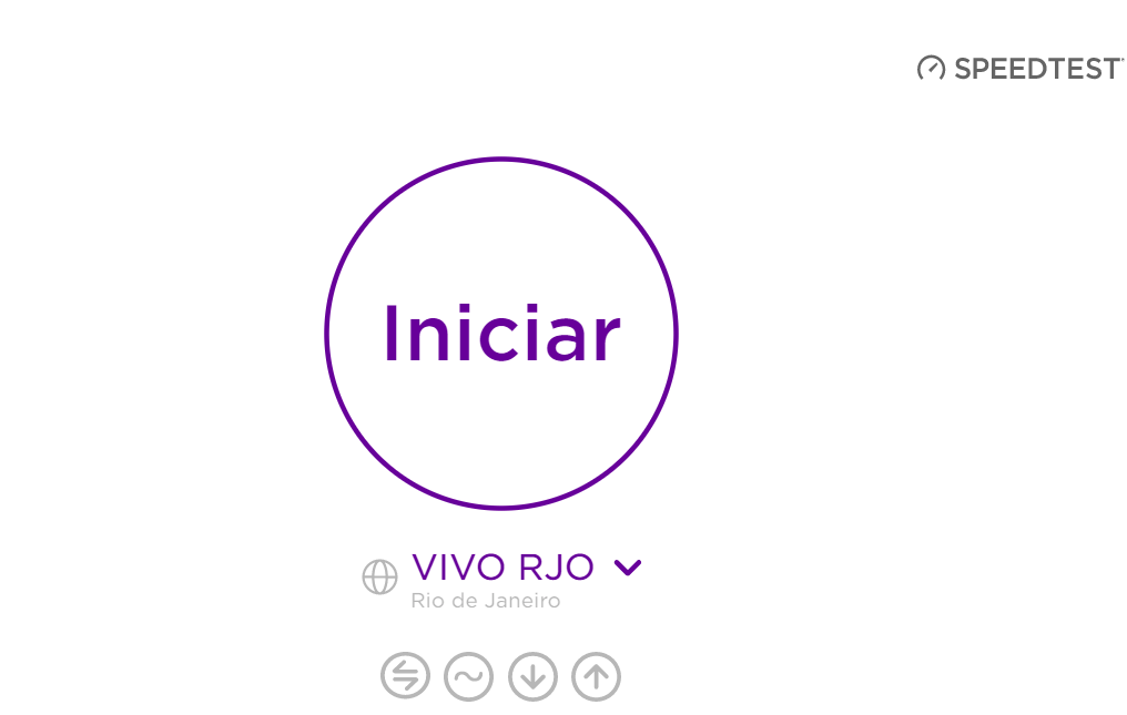 Teste de Velocidade Vivo, Teste Vivo, Power, Internet03 junho 2024
 -
 Teste de internet Vivo Speedy - Medir Conexao03 junho 2024
 -
 Vivo X100 ganha teste com Dimensity 9300 superando Snapdragon 8 Gen 3 - Canaltech03 junho 2024
 -
 Vivo NEX S mostra robustez em teste de durabilidade – Tecnoblog03 junho 2024
 -
 Teste de Velocidade Vivo, Contrate Online03 junho 2024
 -
 Analysis of DMC1 Knockdowns Generated by the In Vivo siRNA03 junho 2024
 -
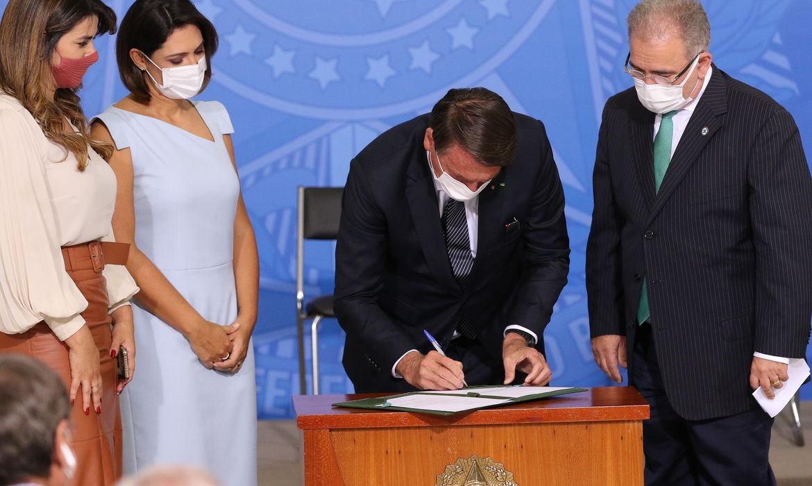 Ao vivo: Bolsonaro sanciona PL que amplia o teste do pezinho03 junho 2024
 -
/i.s3.glbimg.com/v1/AUTH_08fbf48bc0524877943fe86e43087e7a/internal_photos/bs/2022/I/R/Aow0BfRhKUd3kEimLWHw/print2.jpg) Como saber se a Vivo está fora do ar03 junho 2024
Como saber se a Vivo está fora do ar03 junho 2024 -
 Internet movel ilimitada(teste gratis)c3 - Celulares e telefonia03 junho 2024
 -
 Repórter da Record faz teste de Covid-19 ao vivo e resultado dá positivo - ISTOÉ Independente03 junho 2024
você pode gostar
-
 Tiger King': The late-night host who Joe Exotic thinks should play him in a movie03 junho 2024
 -
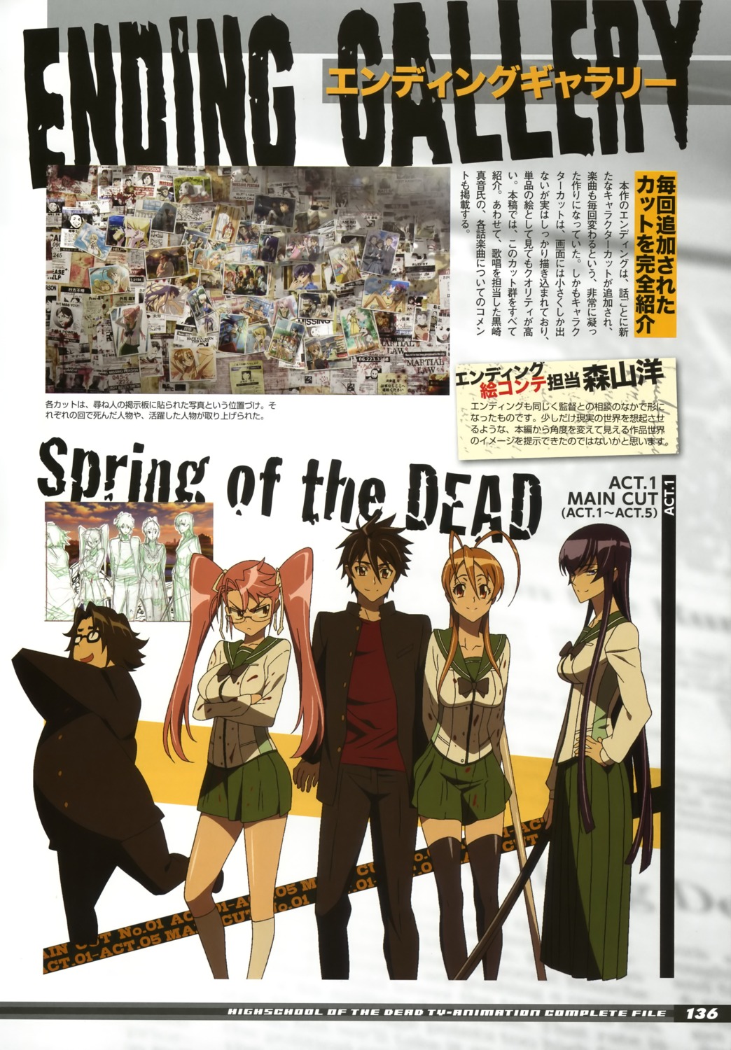 highschool of the dead busujima saeko hirano kohta komuro takashi miyamoto rei takagi saya blood seifuku thighhighs, #18174003 junho 2024
 -
 NARUTO2.o: Nurari03 junho 2024
 -
 What Does an Ascendant or Rising Sign Mean in Your Birth Chart? - Exemplore03 junho 2024
 -
 Evandro Mesquita: Movies, TV, and Bio03 junho 2024
 -
 Bluey Capsules on X: He's starting with the man in the mirror03 junho 2024
 -
 Pack oferta FC Porto - Estadia03 junho 2024
 -
 Ragnarok the Animation (Judia) - Minitokyo03 junho 2024
 -
 Mahjong Classic 2 3.14 Free Download03 junho 2024
 -
 Here's A Great Tony Hawk's Pro Skater 1 & 2 Bargain03 junho 2024