Assessment of Myocardial Viability Using Nuclear Medicine Imaging in Dextrocardia
Por um escritor misterioso
Last updated 03 junho 2024
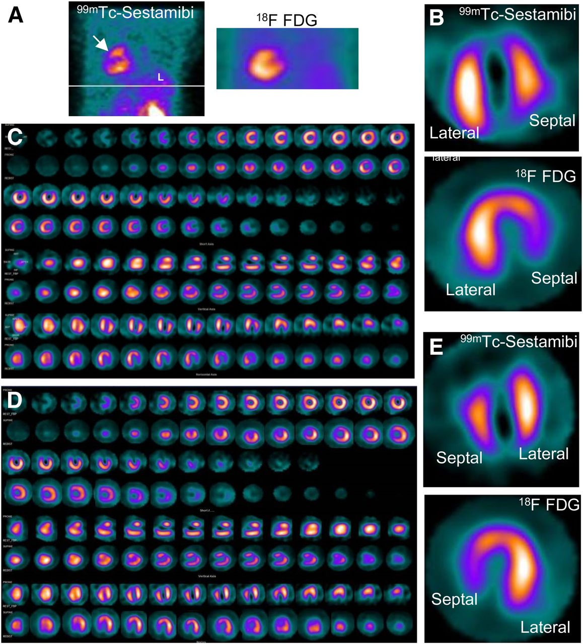
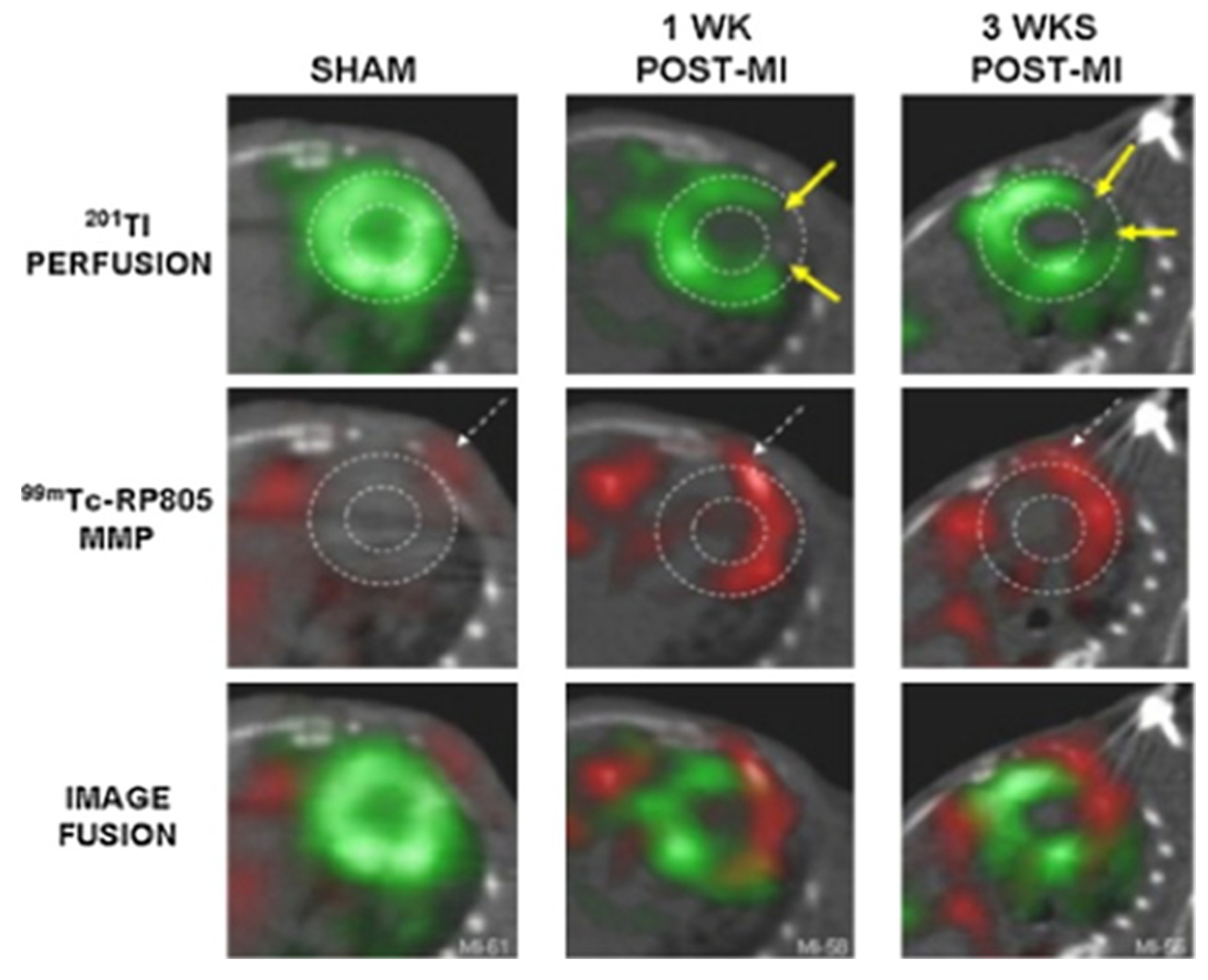
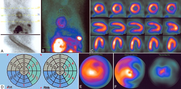
Imaging of dextrocardia in humans requires an understanding of the orientation of the heart chambers and walls. There are many types of cardiac malpositioning, such as dextrocardia (with or without situs inversus), mesocardia, and levocardia. Myocardial perfusion scintigraphy of dextrocardia has been explained in case reports and imaging atlases; however, myocardial viability assessment using nuclear medicine imaging techniques is less documented in the literature. Methods: In 2 cases of dextrocardia with situs inversus and 1 case of mesocardia, myocardial viability was assessed using 99mTc-sestamibi rest perfusion scintigraphy and 18F-FDG PET. Cardiac SPECT images of dextrocardia with situs inversus were acquired using the feet-first supine position with a 180° arc from left anterior oblique to right posterior oblique, whereas a right-lateral–to–left-lateral arc was used for mesocardia. The processing and reconstruction were done by entering the dataset for the feet-first supine position and repeating after entering the dataset for the feet-first prone position. The 2 sets of reconstructed images were compared for orientation of walls and cardiac chambers. Results: The first processing, using the feet-first supine position, revealed an interchanged septum and lateral wall in reconstructed images of dextrocardia with situs inversus. This interchange was corrected by changing the position to prone during processing of the rest perfusion and PET raw data. The display of cardiac slices in various axes matched the conventional nomenclature for the septum and lateral wall, leading to easy interpretation. However, this change was not required in the mesocardia, for which the location of the heart chambers was not interchanged. Conclusion: Because the acquisition protocol for SPECT is a semicircular orbit, the various types of dextrocardia require careful selection of the arc, with the patient positioning kept feet-first supine. Processing and reconstruction of data by changing the patient position to prone was found to be most useful method of matching the septum and lateral wall orientation for interpretation of images.

Viability Studies - Hamilton Cardiology Associates - New Jersey's Leading Board Certified Cardiologists

Myocardial Perfusion and Viability Imaging in Coronary Artery Disease: Clinical Value in Diagnosis, Prognosis, and Therapeutic Guidance - ScienceDirect
Assessing Myocardial Viability in Clinical Practice - ABC Imaging

PET vs MRI for Myocardial Viability - Johann Christopher, 2020
Single Photon Emission Computed Tomography (SPECT) Myocardial Perfusion Imaging Guidelines: Instrumentation, Acquisition, Processing, and Interpretation
Pharmaceuticals, Free Full-Text

Myocardial perfusion single photon computed tomography: An Atlas - ScienceDirect
Pharmaceuticals, Free Full-Text

Motion artifact. Selected NAC and AC SAX images show a markedly

PDF) Myocardial viability assessment using nuclear imaging

What are the 4 components of myocardial viability ? How will you test for it ?

PDF] Myocardial Perfusion SPECT Imaging in Dextrocardia with Situs Inversus: A Case Report

Role of PET-CT in the assessment of myocardial viability in patients with left ventricular dysfunction - ScienceDirect

PDF] Artifacts and pitfalls in myocardial perfusion imaging.
SPECT myocardial perfusion imaging in patients with Dextrocardia
Recomendado para você
-
 Fluid transport in the brain03 junho 2024
 -
 Brain Test Level 372 Walkthrough03 junho 2024
 -
 Brain Test Level 371, 372, 373, 374, 375, 376, 377, 378, 379, 380 Answers03 junho 2024
 -
 Brain Test Nível 372 Respostas - Alejandro03 junho 2024
 -
 Easy Game Brain Test Level 372 Finish shopping.03 junho 2024
 -
 Forty - psychology03 junho 2024
 -
Imaging Surrogates of Disease Activity in Neuromyelitis Optica Allow Distinction from Multiple Sclerosis03 junho 2024
-
Brain Sciences Center - News03 junho 2024
-
 Vessel Part 1-(page 1&2)03 junho 2024
 -
 Autonomy and the principle of respect for autonomy.03 junho 2024
você pode gostar
-
 A Wild Pikamee has appeared! by Towa-MMD on DeviantArt03 junho 2024
 -
 Brain Test Level 191 I hate this! The baby is crying again! Stop this scream Answer03 junho 2024
 -
 Mob Falls in Love in Trailer for Mob Psycho 100 Season 303 junho 2024
 -
RIOCARD DUO APK (Android App) - Baixar Grátis03 junho 2024
-
 adidas Superstar XLG Shoes - White, Men's Lifestyle03 junho 2024
 -
If You're a Taylor Swift Fan, TATPWYFMM Actually Means Something - WSJ03 junho 2024
-
 Exclusivo para PlayStation 4, Horizon Zero Dawn chegará ao PC até03 junho 2024
 -
 World War Z: Aftermath: World War Z: Aftermath Has Arrived on Consoles & PC - Focus Entertainment03 junho 2024
 -
What is now.gg, and how can you play Roblox games on it? - Quora03 junho 2024
-
 Pin em Naruto fotos03 junho 2024